Western Blot Fractions

Anti Ktn1 Antibody Mouse Anti Human Ktn1 Monoclonal Antibody Np 1

Mem Per Plus Membrane Protein Extraction Kit Thermo Fisher Bioz Ratings For Life Science Research

Solved 1 This Image Shows The Results Of A Western Blot Chegg Com
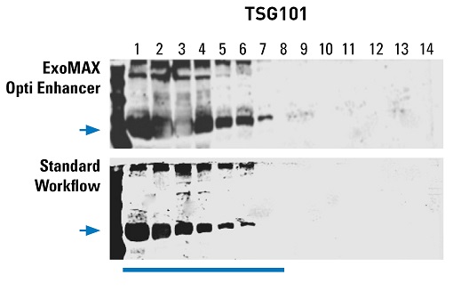
Gradient Based Exosome Isolation

Diagnosis Of Creutzfeldt Jakob Disease By Western Blot Identification Of Marker Protein In Human Brain Tissue Nejm

Western Blot Analysis Of Subcellular Fractions Individual Lanes Download Scientific Diagram
The fractions were examined by western blotting analysis using antibodies directed against specific HK markers and revealed single dense bands for histone H3 at approximately 17 kDa in the nuclear fraction, a band representing GAPDH at approximately 37 kDa in the cytosolic fraction, and a single band at approximately 17 kDa for CoxIV in the mitochondrial fraction (Figure 2 A-C) in each sample.
Western blot fractions. The blot is a membrane, almost always of nitrocellulose or PVDF. I was wondering why this is the case, as Fis1 is mitochondrial protein, and if it would be appropriate to western blot for Fis1 in the mitochondria fraction alone to increase signal. WESTERN BLOTTING - A BEGINNER’S GUIDE Western blotting identifies with specific antibodies proteins that have been separated from one another according to their size by gel electrophoresis.
Moreover, western blot analysis and Caspase GloTM luminescent assay demonstrated that fractions F2 and F3 triggered apoptotic cell death via activation of caspases -8, -9 and -3/7 and up-regulation of Bax and down-regulation of l-2 protein. Thermo Fisher Scientific's Invitrogen cell lysis and organelle analysis kits are optimized for stepwise separation, enrichment, and extraction of proteins from different cell fractions, including cytoplasmic, membrane, nuclear, chromatin-bound, and cytoskeletal proteins in 1–3 hours. Western blot trouble shooting, problems with western blot, western trouble, western blocking time, blotting time, primary antibody detection, incubation with primary antibody, membrane storage, sample extraction, western blot for plant and algal samples.
Also used as a nutrient in cell and microbial culture. Load 10 μg each of the cytosolic and mitochondrial fractions isolated from uninduced and induced cells on a 12% SDS-PAGE. Ponceau S staining solution:.
Experiment 7 – Lab Report. Nuclear RIPA or use nuclear fraction protocol*. Place the tissue in round-bottom microcentrifuge tubes or Eppendorf tubes and immerse in liquid nitrogen to snap freeze.
MM Tris-HCl, pH 7.5, 150 mM NaCl, 5% (w/v) skimmed milk powder, 0.02% sodium azide, 0. Add up all primaries (non-conjugated, 100% specific) and secondaries. It is a microsomal ascorbate peroxidase, which scavenges hydrogen peroxide in plant cells.
HRP conjugated secondary antibody cocktail at 1/2500 dilution Predicted/observed band sizes:. • Buffer component in immunochemistry, biochemistry, cell biology, or molecular biology • Reducing agent. Unboiled samples or special gel systems).Please refer to the remarks sections for western blotting on the respective data sheet.
Western blot analysis Equal amounts (based on protein assay) of total protein (10µg or 30µg) were resolved on denaturing 4-% Tris-glycine SDS-polyacrylamide gels and then transferred to nitrocellulose membranes. The positive bands from both my first and second IgM tests were 18, 30, 31, 34, and 41. ARG anti-GAPDH antibody 6C5 WB image.
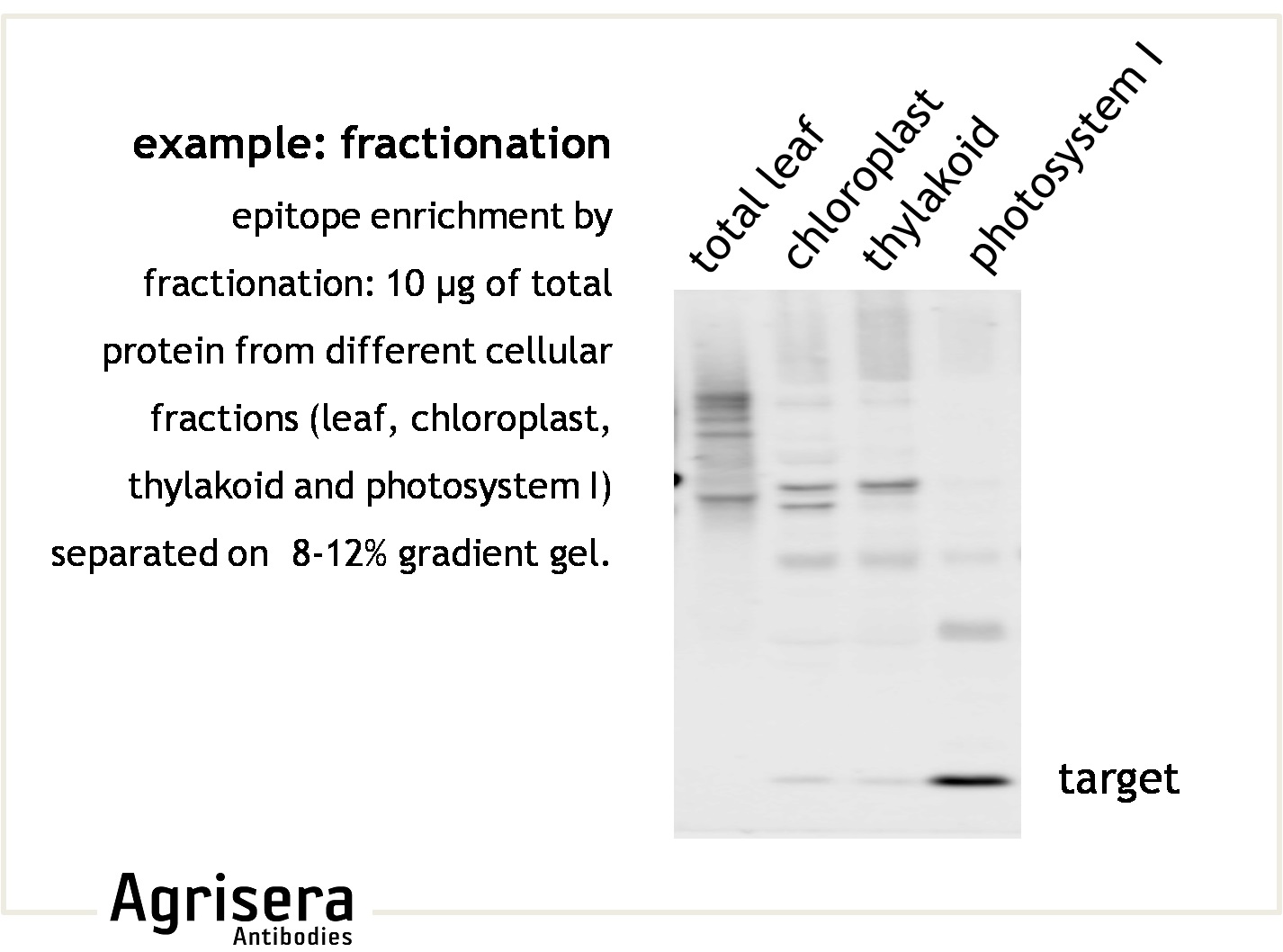
Some proteins have special requirements for good separation (e.g. If you are carrying out Western blotting on individual fractions, or organelles, it is important that you can confirm you are looking at the correct organelle fraction. In 1979, Jaime Renart et al.
Published an article entitled “ Transfer of proteins from gels to diazobenzyloxymethyl-paper and detection with antisera:. Relative Activity (Units/mL) F1 0 0 F2 0.002 0.096 F3 0.093 0.748 F4 0.053 2.556 F5 0.045 2.170 F6 0.016 0.772 F7 0 0 F8 0 0 F9 0 0 F10 0 0 F11 0 0 F12 0 0. Experiment 7 – Lab Report Western Blot Analysis of rGFP fractions.
Bands 23-25 and 39 were reported as indeterminate in the first IgM WB but only band 39 showed up as indeterminate in the second IgM WB. Based on the system discussed in class, what is the minimal number of different antibodies that had to be used to generate this western blot?. Western blotting, also known as immunoblotting, is a well-established and widely used technique for the detection and analysis of proteins.The method is based on building an antibody:protein complex via specific binding of antibodies to proteins immobilized on a membrane and detecting the bound antibody with one of several detection methods.
Before transferred proteins can be detected using antibodies, the membrane must be incubated with a blocking buffer to block the unoccupied binding surfaces on the membrane. Unboiled samples or special gel systems).Please refer to the remarks sections for western blotting on the respective data sheet. Western blotting is a very common and powerful technique often used worldwide to detect, characterize and quantify proteins.
RIPA buffer is possibly one of the most commonly used cell lysis buffers for detection of phosphorylated protein by Western blot. Western Blot Analysis of rGFP fractions. Rapid Preparation of a Plasma Membrane Fraction:.
• Stabilizer to prevent proteolysis and denaturation of enzymes and other proteins, especially when the proteins are present in low concentrations. Dissect the tissue of interest with clean tools, on ice preferably, and as quickly as possible to prevent degradation by proteases. Although common, a Western blot is composed of multipl.
Lanes 2,4 & 6 represent supernatants (soluble fractions) from jas-plakinolide treated cell lysates. <p>(A) The ability of the different antibody fractions to detect a protein of the expected molecular weight in Western blot is shown in green (positive) or red (negative). 78 kDa / 75 kDa ATP5A band size:.
This is the Cytoskeletal and Nuclear Fraction. Luckily antibody companies like Proteintech have many organelle- or fraction-specific control antibodies (see below), which can be used to verify that the isolated cell component you’re looking at is the right one. Coli strain BL21(D)<pLysS><pRSETA-GFP UV > using the Ni+2.
Western blot analysis can be used to confirm enrichment by probing a protein whose expression is restricted to the subcellular fraction of interest (e.g. It helps to identify the presence of a specific protein both by size and through the binding of an antibody makes them well-suited for evaluating levels of protein expression in cells, and for monitoring fractions during protein purification. Why is RIPA Buffer Best for Western Blot?.
A) In which fractions does the DNA binding activity reside (N/E represents nuclear extract (the starting material) and (-) is the reaction performed with buffer-no protein. When investigating plasma or serum by Western blotting, abundant plasma proteins, such as albumin and IgG can obscure the signals of less abundant proteins. In brief, the sample undergoes protein denaturation, followed by gel electrophoresis.A synthetic or animal-derived antibody (known as the primary antibody) is.
Load 15 µl of each fraction along with 15 µl of WCL. Explain your answer in 2 sentences maximum. Select the right subcellular fractionation kit for you.
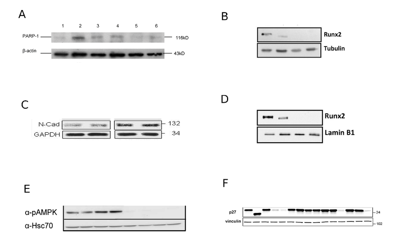
5% acetic acid, 0.1% Ponceau S 5% skimmed milk-TBST:. Antigenic fractions of 100, 50, 37, and 28 kDa obtained through the SDS-PAGE method that were more frequently recognized by anti-Coccidioides antibodies in the sera of coccidioidomycosis patients were selected using western blotting.Subsequently, these bands were sequenced, and the obtained proteins were analysed by BLAST to choose peptides specific for Coccidioides spp. The nuclear fraction should demonstrate more robust Lamin B1 signal than the cytoplasmic fraction following nuclear/cytoplasmic fractionation.
The number indicates the relative amount in percent of each antibody fraction and a total of antibody fractions towards linear epitopes that are able to bind the target protein in Western blot. Then proceed with a standard Western blot procedure and probe with the cytochrome c antibody (recommended working concentration is 1 μg/ml) and the proper organelle markers. Ponceau S staining solution:.
Run the immunoprecipitated fraction on a gel and then perform a Western blot using the modification-specific primary antibody. Membranes were blocked with 5% BSA in Tris-buffered saline with Tween™ for 1 hour. In Arabidopsis there are three cytosolic (APX1, APX2, APX6), two chloroplastic types (stromal sAPX, thylakoid tAPX), and three microsomal (APX3.
5% acetic acid, 0.1% Ponceau S 5% skimmed milk-TBST:. Lanes 1,3 & 5 represent pelleted fractions from jasplakinolide treated cell lysate. Western blot analysis of γ-synuclein in protein fractions obtained by sequential extraction of the dorsolateral column area samples of sALS patients with (+ve) or without (−ve) γ-synuclein.
Membrane Fraction Western blot cocktail at 1/250 dilution Secondary antibody:. Bovine Serum Albumin (BSA) is used for various biochemical applications including ELISA (Enzyme-Linked Immunosorbent Assay), high content screenining assays, western blotting, and immunohistochemistry. A method for studying antibody specificity and antigen structure,” the prelude to the modern Western blot (WB) technique.
The ELISAs were performed according to the manufacturer's instructions. The input (I) and the co-immunoprecipitated fraction (B) of the RFP-labeled proteins were being visualized via western blot using anti RFP antibody Western blotting was executed as described prior to 33 transferring the proteins on a nitrocellulose membrane (GE Health care, Munchen, Germany). Membrane proteins account for 70% to 80% of all pharmaceutical targets, indicating their clinical relevance and underscoring the importance of identifying differentially expressed membrane proteins that reflect distinct disease properties.
Western Blot Analysis of rGFP fractions BIOL3380 - Fall. Rapid Preparation of a Plasma Membrane Fraction:. Yamamoto N(1), Yamashita Y(2), Yoshioka Y(3), Nishiumi S(4), Ashida H(2).
2.3.2 Results and discussion. Boil each sample for 5 min at 95°C and centrifuge for 3 min at 15,000 x g. Lamin B1 is expressed in the nucleus, but not the cytoplasm).
Fraction Δ Abs/ Δ min. Went one step further and published. Sodium Potassium ATPase = 112 kDa/100 kDa GRP78 band size:.
It is recommended to confirm the enrichment of subcellular extracts following isolation. Western Blot Controls Overexpressed Membrane Fractions Voltage-dependent Ca 2+ channels (Ca V ) form an important route for Ca 2+ entry into cells, upon deviations from the cells’ resting membrane potential. Purification of LDH Homogenization, centrifugation, and ammonium sulfate precipitation.
A major problem with Western blotting is not the method itself, but the use of poor quality antibodies as well as the use of different experimental conditions that affect the linearity and sensitivity of the Western blot. Western Blot for Endoplasmic Reticulum Membrane Antibody Cocktail – Cell Fractionation HeLa cell lysates were prepared using the Membrane Fractionation Kit ( ab ), blots were developed using the ECL technique, performed under reducing conditions and exposed for 5 minutes. The western blot (sometimes called the protein immunoblot), or western blotting, is a widely used analytical technique in molecular biology and immunogenetics to detect specific proteins in a sample of tissue homogenate or extract.
However, too stringent elution conditions may result in large amounts of antibody being eluted off the beads and western blot detection of these antibodies, specifically their denatured forms (the antibody heavy and light chains seen on western blots of IP samples originate from the antibody used during the IP procedure). Western Blot Detection of Translocated Glucose Transporter 4 from Plasma Membrane of Muscle and Adipose Cells and Tissues. Western Blot with anti-LDH antibody.
Investigation of some conditions that are commonly used and often modified in Western blotting, as well. Soon after, Harry Towbin et al. Bovine Serum Albumin Fraction V is suitable for standard applications such as:.
Sonicate for 5 sec at % power 3 times. Prepacked columns, such as HiTrap™ Albumin & IgG Depletion are designed to deplete samples of these potentially problematic proteins, removing >95% albumin and >90% IgG, respectively. Experiments #3-7 The overall goal/purpose of this series of experiments is to see if we can express and purify a His 6-tagged recombinant form of GFP (rGFP) from the E.
Membranes used for Western blotting have a high affinity for proteins allowing binding and retention of transferred proteins. Western Blot Detection of Translocated Glucose Transporter 4 from Plasma Membrane of Muscle and Adipose Cells and Tissues. For western blot Preparation of lysate from tissues 1.
The following gel of various unrelated proteins was used for a western blot. A Western blot provides a readout of differences in protein expression levels e.g. Supernatant (S) and pellet (P) fractions and analyzed by western blot quantitation of actin protein accord-ing to the G-actin/F-actin In Vivo Assay Kit instruc-tions.
Chemical treatments to remove modifications Proteins and protein extracts can be treated with chemicals that will remove modifications (e.g. From among the shared. Some proteins have special requirements for good separation (e.g.
My own IGeneX Western Blots were both positive by IGeneX criteria but negative by CDC criteria. 60 kDa / 60 kDa GAPDH band size:. Simply edit this word document by inserting your answers after each question/space.
Densitometric analysis of the Western blots indicated that the linear range for IκBα was 0.097–3.12 ng and for p53 was 1.87. PNGase F is used to removed glycosylations). The quantitative Western blot data were confirmed using p53 and IκBα ELISA assays (Assay Designs, Ann Arbor, MI).
MM Tris-HCl, pH 7.5, 150 mM NaCl, 5% (w/v) skimmed milk powder, 0.02% sodium azide, 0. (1)Research & Development Institute, House Wellness Foods Corporation, Itami, Japan. (Give fraction numbers) B) How does association with other proteins affect the ability of this protein to bind DNA?.
Western Blot Controls Overexpressed Membrane Fractions AMPA receptors are members of the glutamate receptor family of ion channels that also include the NMDA and Kainate receptors. BSA as a blocking reagent is particularly useful with casein-sensitive antibodies, such as phospho-specific antibodies. Western Blot Applications Western blot technique has a wide range of applications in scientific and clinical disciplines.
Western blotting is a commonly used technique in biological research. Store samples at -80°C for later. APX3, ascorbate peroxidase 3, has been used as a Western blot loading control for plant membrane fractions.
Add 60 μl of 3X SDS Loading Buffer with DTT (#7722) for every 100 μl of supernatant. Polyclonal antibodies (serum or IgY-fractions from egg yolk) usually contain a number of. Western Blot Analysis of rGFP fractions.
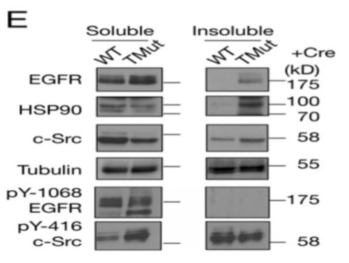
37 kDa / 38 kDa. 1) U87-MG 2) MCF-7 3) A549 4) DU145 5) SW480 6) rat brain 7) rat stomach 8) rat ovary stained with ARG anti-GAPDH antibody 6C5 at 1:5000 dilution. RIPA soluble and RIPA insoluble fraction.
Detection Of Mclk1 In Protein Fractions Western Blot Analysis Of Download Scientific Diagram
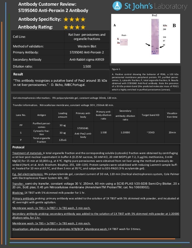
Western Blot Antibody Customer Review For Anti Peroxin 2 Antibody St

Pin On Chemistry

Smn Affects Membrane Remodelling And Anchoring Of The Protein Synthesis Machinery Journal Of Cell Science
Western Blot Of Nuclear And Cytosolic Fractions Of The Cancer Cell Download Scientific Diagram
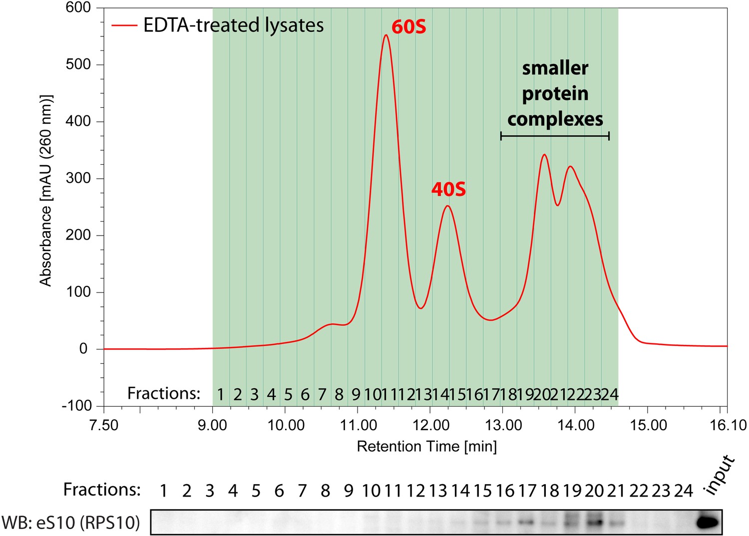
Efficient Analysis Of Mammalian Polysomes In Cells And Tissues Using Ribo Mega Sec Elife
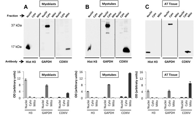
A Simple Protocol For The Subcellular Fractionation Of Skeletal Muscle Cells And Tissue Springerlink

Functional Analysis Of Hif1 Histone Chaperone In Saccharomyces Cerevisiae G3 Genes Genomes Genetics
Loading Controls For Western Blots
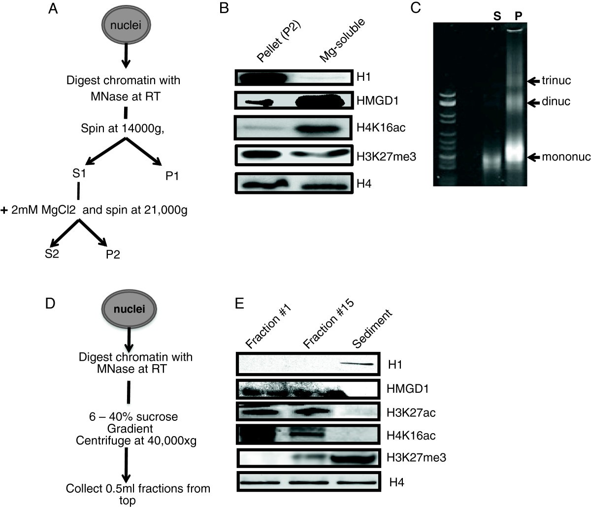
The Chromatin Architectural Proteins Hmgd1 And H1 Bind Reciprocally And Have Opposite Effects On Chromatin Structure And Gene Regulation Bmc Genomics Full Text
Solved Question 32 1 Pts If You Are Presented With A Grap Chegg Com
A Simple Protocol For The Subcellular Fractionation Of Skeletal Muscle Cells And Tissue Springerlink

Figure 2 Western Blot Analysis Of The Ptm Complex Containing Fraction

Pdf Western Blot Antibody Determination In Sera From Patients Diagnosed With Anisakis Sensitization With Different Antigenic Fractions Of Anisakis Simplex Purified By Affinity Chromatography Semantic Scholar

Sds Page And Western Blotting Of The Isolated Fractions

Team Ucopenhagen Notebook Week 42 19 Igem Org
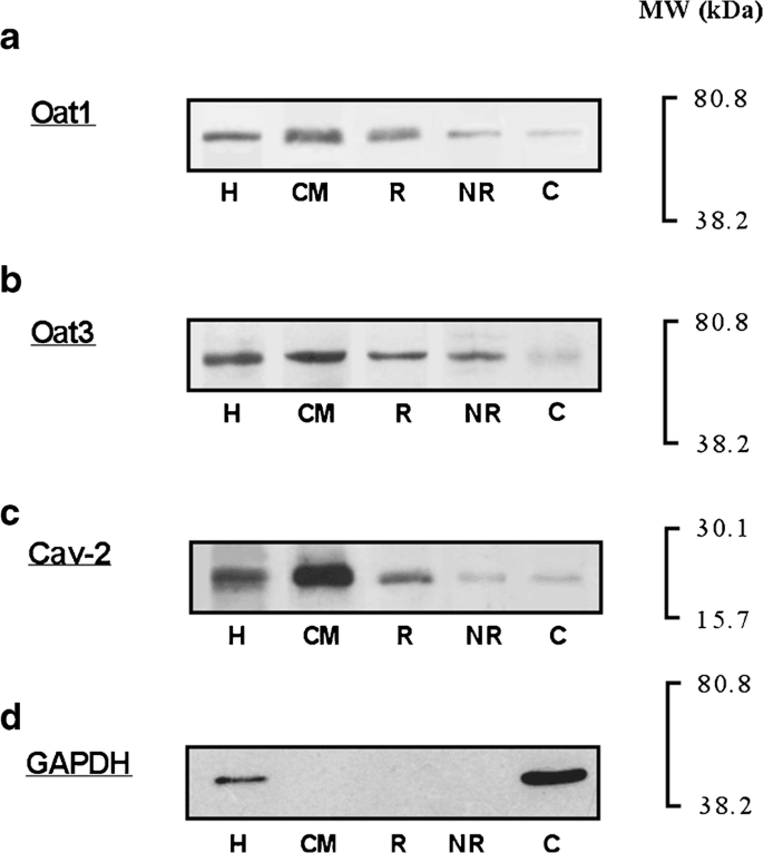
Distribution Of The Organic Anion Transporters Oat1 And Oat3 Between Renal Membrane Microdomains In Obstructive Jaundice Springerlink

Affinity Purification Chromotek
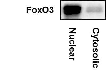
Foxo3 Antibody Nbp2 Novus Biologicals
Q Tbn 3aand9gcrd Hgcule2fv1gd9icxftgqxkk8knqgmae5ldx3zqbjtru3 Usqp Cau
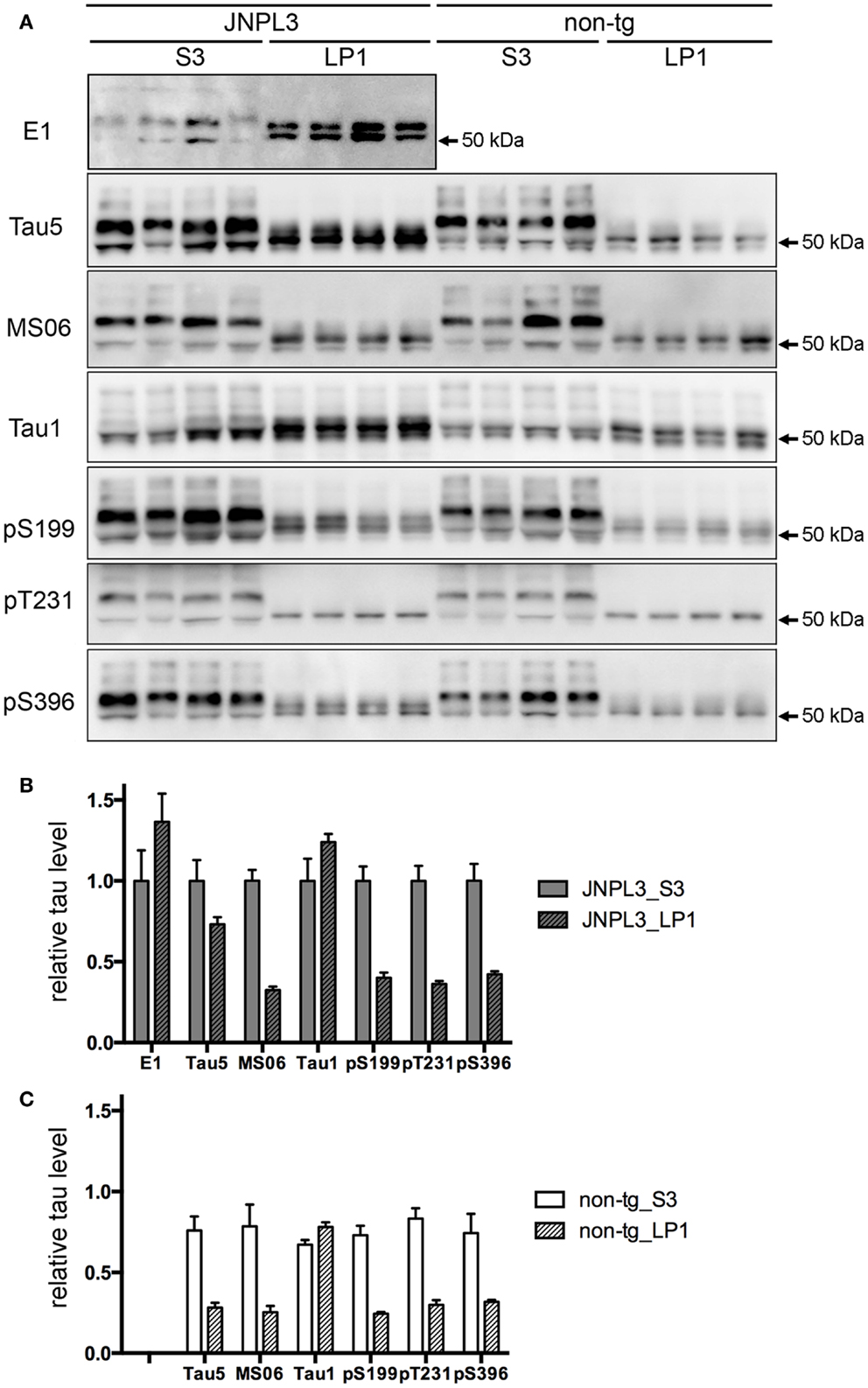
Frontiers Biochemical Distribution Of Tau Protein In Synaptosomal Fraction Of Transgenic Mice Expressing Human P301l Tau Neurology
Western Blot

Lysosome Fraction Western Blot Cocktail Ab Abcam
Insight Jci Org Articles View 590 Sd Pdf Render 1

Anti P35 Cdk5 Regulator Antibody Produced In Rabbit Igg Fraction Of Antiserum Buffered Aqueous Solution Sigma Aldrich

Agrisera Western Blot Recommendations Troubleshooting And Protocol
Sds Page And Western Blot Analysis Of Recombinant Cedna Open I

Western Blot Analysis Of Polyribosome Fractions Of Hcv Ires Gfp Transfected Cells

Visualization Of Protein Protein Interaction In Nuclear And Cytoplasmic Fractions By Co Immunoprecipitation And In Situ Proximity Ligation Assay Protocol
Plos One B Cop As A Component Of Transport Vesicles For Hdl Apolipoprotein Mediated Cholesterol Exocytosis

Analysis Of Lap Distribution In Cellular Fractions A Western Blot Download Scientific Diagram
Western Blot Analysis Of N Protein In Nuclear And Cytoplasmic Fractions Download Scientific Diagram
2

Western Blot Sample Preparation Sino Biological

A Western Blot Analysis Of Membrane Fraction Obtained From 6 Week Old Download Scientific Diagram
Q Tbn 3aand9gcs2uae4rralkx2rbjubn8ytmafhoujza6j0siwe Iztbatfzfq3 Usqp Cau

Western Blot Analysis On Cellular Proteins Separated Into Nuclear And Cytoplasmic Fractions From Snf96 2 Cells

Differential Fractionation Of Erythrocytes Infected By
-Western-Blot-NBP2-22190-img0006.jpg)
Foxo1 Fkhr Antibody 3b6 Nbp2 Novus Biologicals

Membrane Fraction Wb Cocktail Ab Abcam

Isolation Of Pure Mitochondria From Rat Kidneys And Western

Anti Vp16 Antibody Produced In Rabbit Igg Fraction Of Antiserum Buffered Aqueous Solution Sigma Aldrich

Lysosome Fraction Western Blot Cocktail Ab Abcam

Silver Staining And Western Blot Analysis Of Blue Pass Fraction Treated With Or Without Glycosidase
Western Blot Analysis Of Cytoplasmic And Nuclear Protein Fractions In Download Scientific Diagram
Www Bio Rad Antibodies Com Static 16 Cancer 10 Tips For The Western Blot Detection Of Ip Samples Pdf

Functional Analysis Of Hif1 Histone Chaperone In Saccharomyces Cerevisiae G3 Genes Genomes Genetics

Figure 3 From Human Alkb Homolog 1 Is A Mitochondrial Protein That Demethylates 3 Methylcytosine In Dna And Rna Semantic Scholar

Diagnosis Of Creutzfeldt Jakob Disease By Western Blot Identification Of Marker Protein In Human Brain Tissue Nejm
Phosphoproteins By Western Blot Stratech

The Molecular Chaperone Hsp40 Regulates The Activity Of P58ipk The Cellular Inhibitor Of Pkr Pnas

Mitochondria Fraction Western Blot Cocktail Ab Abcam
Phosphoproteins By Western Blot Stratech
Www Unco Edu Nhs Biology About Us Pullen Nicholas Documents Pullen Lab Protocol For Western Blotting Transblot Pdf

Western Blot Analysis Of The Plasma Membrane Fraction Where Cadherin Download Scientific Diagram

Vdac1 Antibody Nb100 695 Novus Biologicals

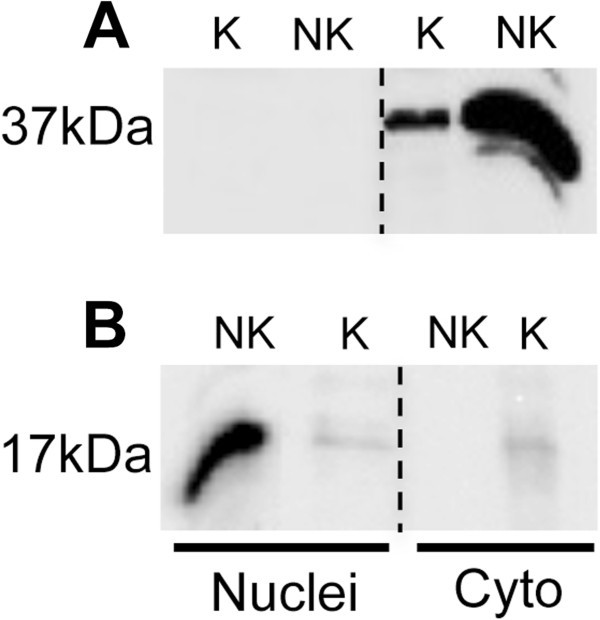
Part a K Parts Igem Org
High Resolution Preparation Of Monocyte Derived Macrophages Mdm Protein Fractions For Clinical Proteomics Proteome Science Full Text
Microsome Isolation Kit Ls K159
Ataxia Telangiectasia Mutated Is Located In Cardiac Mitochondria And Impacts Oxidative Phosphorylation Scientific Reports
Western Blot Analysis Of Oocytes Expressing Wt And S6 M Open I

Plasma Membrane Fraction Western Blot Cocktail Ab Abcam

Proteome Phosphoproteome And Hydroxyproteome Of Liver Mitochondria In Diabetic Rats At Early Pathogenic Stages Molecular Cellular Proteomics

Figure 10

Western Blotting Analysis Of The Gfap Fragments In The Soluble And Insoluble Protein Fractions Of Als And Non Als Spinal Cords
Frontiers Biochemical Distribution Of Tau Protein In Synaptosomal Fraction Of Transgenic Mice Expressing Human P301l Tau Neurology

Cell Fractionation Kit Standard Ab Abcam
An Optimized Procedure For Isolation Of Rodent And Human Skeletal Muscle Sarcoplasmic And Myofibrillar Proteins Roberts Journal Of Biological Methods

Gfp Trap Chromotek
Western Blot Analysis Of Retinal Protein Fractions A Glucose Download Scientific Diagram

Spot Trap Chromotek

Caspase 3 Antibody Western C9598 Sigma Aldrich
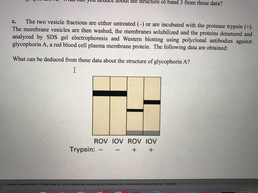
Solved The Two Vesicle Fractions Are Either Untreated Chegg Com

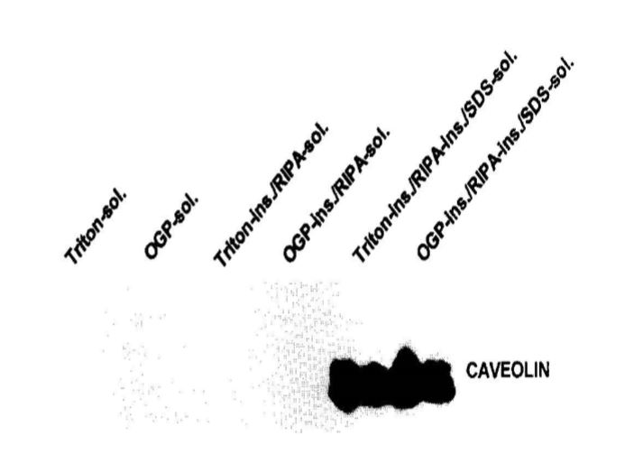
Alterations In Detergent Solubility Of Heterotrimeric G Proteins After Chronic Activation Of Gi O Coupled Receptors Changes In Detergent Solubility Are In Correlation With Onset Of Adenylyl Cyclase Superactivation Molecular Pharmacology

Anti Groes Antibody Produced In Rabbit Igg Fraction Of Antiserum Buffered Aqueous Solution Sigma Aldrich

Anti Ef1a3 Antibody Mouse Anti Human Ef1a3 Monoclonal Antibody Q5vte0 1
Experimental Design Strategy

Lc3 Ii Enrichment Cancer Life Science Research Emd Millipore

Figure 2 From Mdr Tb Antibody Response Western Blot To Fractions Of Isoniazid And Rifampicin Resistant Antigens Of Mycobacterium Tuberculosis Semantic Scholar
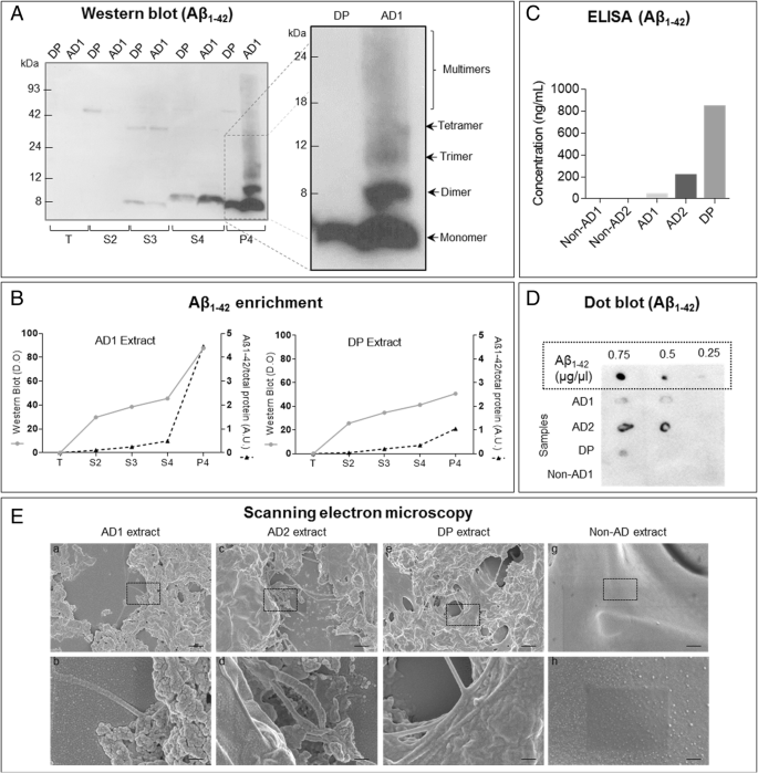
Human Amyloid B Enriched Extracts Evaluation Of In Vitro And In Vivo Internalization And Molecular Characterization Alzheimer S Research Therapy Full Text

Cytosol Markers Abcam Bioz Ratings For Life Science Research

Jci Insight Intracellular Localization Of Diacylglycerols And Sphingolipids Influences Insulin Sensitivity And Mitochondrial Function In Human Skeletal Muscle

A Method To Separate Nuclear Cytosolic And Membrane Associated Signaling Molecules In Cultured Cells Science Signaling
Plos One Sirt1 Activity Is Linked To Its Brain Region Specific Phosphorylation And Is Impaired In Huntington S Disease Mice

Endoplasmic Reticulum Fraction Western Blot Cocktail Ab Abcam
Ijms Free Full Text Tau Fibril Formation In Cultured Cells Compatible With A Mouse Model Of Tauopathy Html
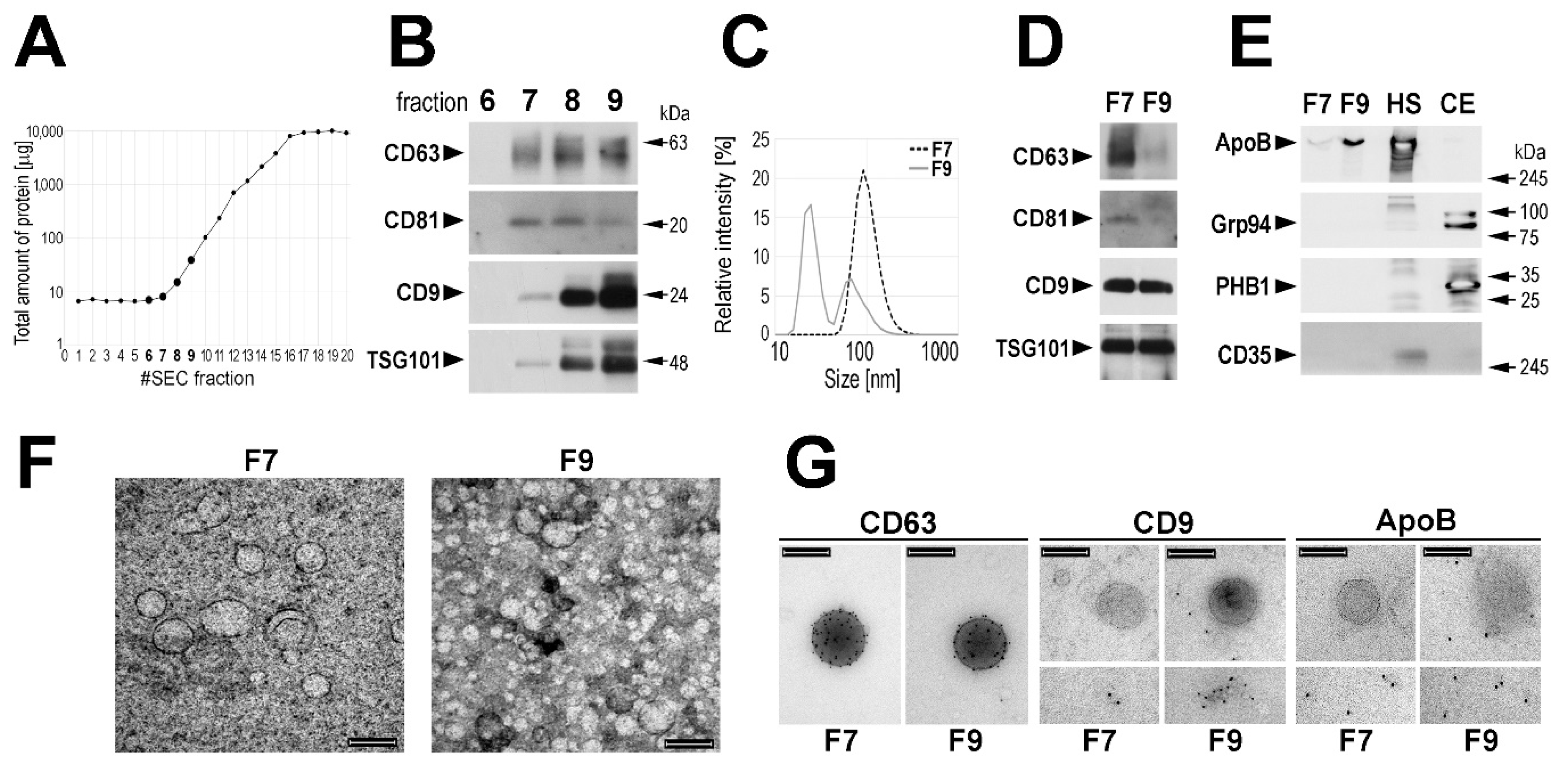
Proteomes Free Full Text Proteome Profiling Of Exosomes Purified From A Small Amount Of Human Serum The Problem Of Co Purified Serum Components Html
Q Tbn 3aand9gcskhjbewghy05hrzxgefnoqykdeue5uojku 0dkaixjlikpz3u5 Usqp Cau

Trek 1 Antibody Nb110 Novus Biologicals
Western Blot Analysis Of Size Fractions Of Membrane Com Open I
Plos One Subcellular Fractionation Analysis Of The Extraction Of Ubiquitinated Polytopic Membrane Substrate During Er Associated Degradation

Team Ucopenhagen Notebook Week 42 19 Igem Org

Mitochondrial Hmgb1 Is Linked To Mitochondrial Functions Western Blot With Separated Cellular Components From The Cerebellar Tissues Of The Three Genotypes Ppt Download

Close Fig 5 Nuclear Shuttling Of Nfatc1 And Epigenetic Histone H3 Modifications Upon Acute Exercise A Confirmation Of Nuclear And Sarcoplasmic Fractions In Skeletal Muscles We Performed Western Blot For A Nuclear Marker Protein Total H3 And A

Figure 2 Detection Of Psvs In B A B Dot Blots Of Fractions 6 14 B B B L1 Western Blots Of Fractions 8 13 B C B L2 Western Blots Of Fractions 8 13 And B D B Electron Micrographs Of Hek293tt
Western Blotting Of Nuclear And Cytosolic Fractions Of Ovcar 3 Cells Download Scientific Diagram
Q Tbn 3aand9gcq1uriwvelidcsfvwkqhvidbxeluqgjh6d5agucqgmolhbc3t Usqp Cau

Anti Roa1 Antibody Mouse Anti Human Roa1 Monoclonal Antibody Jao 1


