Protein Assay Graph

Assays For Determination Of Protein Concentration Olson 07 Current Protocols In Protein Science Wiley Online Library

Figure 4 From Linearization Of The Bradford Protein Assay Increases Its Sensitivity Theoretical And Experimental Studies Semantic Scholar
Solved Calculate The Protein Ug Tube From The Graph Belo Chegg Com

Assays For Determination Of Protein Concentration Olson 07 Current Protocols In Protein Science Wiley Online Library

Protein Quantitation

Bradford Assay Kit Ab Abcam
To analyze data from standard curve and unknowns.

Protein assay graph. Unfortunately, this assay isn’t compatible with lots of common chemicals:. In some research applications, this assay is recommended as a replacement for other protein assays, especially the widely used. 0.04mg 1mg BSA protein used the linear part of the curve.
0.005mg t 0.050mg BSA protein used, the linear part of the curve. To know determine the actual. The protein sample, irrespective of its composition, is measured through absorbance spectroscopy at 540 nm in conjunction with a k.
-Graph known amount of protein vs. The standard curve and the unknowns. Page 6 of 8.
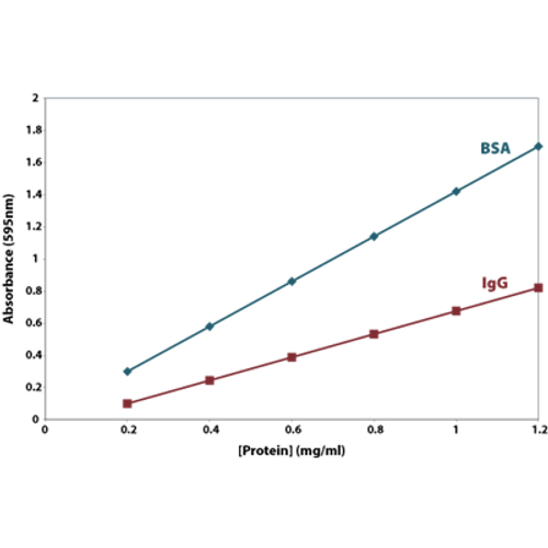
About twice as sensitive to BSA as to IgG. Absorbance and amount of protein are continuous variables, so we should add a trend line that relates absorbance to amount over the entire usable range of the assay. Validation involves a risk assessment step that is followed by a close analysis of the robustness of each assay.
Unlike BCA, this is an endpoint assay with a stable result, meaning that you can estimate the amount of protein from one assay by comparing it with a previous standard curve!. The key component in this assay is bicinchoninic acid (BCA) which reacts with cuprous ions to generate an intense purple color at 562 nm. For greater accuracy run this step in duplicate.
The Bradford is recommended for general use, especially for determining protein content of cell fractions and assesing protein concentrations for gel electrophoresis. How does this absorbance relate to the actual protein concentration?. 5000 microplate assays.
Slightly exceed the standard concentration of gelatin. To prepare a standard curve. While doing a protein assay throughout this semester.
The reagent Coomassie Brilliant Blue turns blue when it binds to arginine and aromatic amino acids present in proteins, thus increasing the absorbance of the sample. Reference to absorbances obtained for a series of standard protein dilutions, which are assayed alongside the unknown samples. To pipet accurately.
How the Bradford Protein Assay Works. • Qualitative.An ELISA can be used to determine whether the protein of interest is found within a particular sample by comparing the sample to a blank well or a sample that does not. Linearity of the assay.
You are aware that A 280 is about twice as sensitive to IgG as to BSA. Note that the equation describing the regression curve is provided along with statistics concerning the curve. Plotting a graph with the absorbance value as the dependent variable (Y-axis) and concentration as the independent variable (X-axis), results in an equation formatted as follows:.
Bradford assay is a protein quantification protocol developed by Marion Mckinley Bradford in 1976. A protein assay consists of two main components:. Standard Curve The Bradford assay is calibrated by using a pure protein of known concentration, called the "standard protein".
Calibration curve for BSA standard is prepared using standard albumin, 50 ml Pierce with concentration of 2 g/l, diluted with 1 M NaOH solution. The dye reagents are commonly purchased from Bio-Rad (Richmond, CA). The Lowry protein assay method for protein concentration determination is one of themost venerable and widely-used protein assays.
The subcellular fractionation of the cauliflower, 2. The Biuret Assay, also known as the Piotrowski Test, is a biochemical assay that allows one to accurately quantify protein concentration within the range of 5-150 mg/mL. When the Bradford reagent (acidified Coomassie Brilliant Blue G-250) binds to proteins, the dye undergoes a color change in the visible spectrum, with the absorbance maximum moving from 470 to 595 nm.
Example of Assay Data Table Create a tablewith the absorbance results obtained from the assay. Stock of Bradford reagent. The enzyme-linked immunosorbent assay (ELISA) is a sensitive technique for determining the concentration of a protein in a liquid sample.
Bradford and Lowry protein assay reagents results in a change in absorbance when protein is present. Explain why or why not. When the dye is allowed to interact with a substance that contains protein, the arginine groups of the protein bind to Coomassie.
The BCA Protein Assay has been advertised as an alternative to the Lowry assay. Absorbance was measured at 750 nm using visible spectrophotometry, using 0.4 mg/mL BSA as the protein standard. This is if the standard curve is linear, however, with BSA standard curve (x axis is time starting from 0) it is not linear and is not that simple.
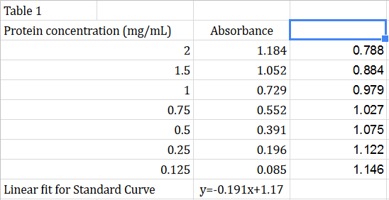
The Bradford assay relies on the binding of the dye Coomassie Brilliant Blue G250 to protein, in which the dye is proportional to the protein concentration. Revised Fall 10 Biuret Protein Assay. (absorbance) for the standards → we are going to create a standard curve and determine the slope of the line.
Using standard procedure, the assay is used with samples having protein concentrations between 0 and 1,400 µg/ml (–140 µg total). Biuret Protein Assay - based on binding of copper ions to peptide bonds under. # BE -402C ).
In assays using 5 ml color reagent prepared in the lab, the sensitive range is closer to 5 to 100 µg protein. Read the absorbance with a spectrophotometer at 595 nm. Reagents and equipment needed:.
The Bradford protein assay is a time-tested colorimetric assay. When performing ELISAs, three different data outputs can be generated:. Standard curve prepared using the protein standards (Table 2 and Graph 1).
And i do get R square (R2)= 0.97 which is closer to 1. When you graph your protein assay data in Excel, should you include the absorbance of your unknown BSA sample?. Asked Sep 18, 19.
This assay works by measuring the color change achieved with the basic amino acids combined with Coomassie dye, which, under. View the graph that is produced from the data by clicking Protein Standard Curve under Graphs in the navigator bar. Depending on the sample type and purpose for performing an assay, protein-to-protein variation is an important consideration in selecting a protein assay method and in selecting an appropriate assay standard (e.g., BSA vs.
I did Bradford assay to get a standard curve in order to determine the protein concentration. Bradford Protein Assay - based on the binding of prot ein molecules to. As each protein requires a tailored quantification method this will to some extent.
From the graph, the protein concentration in raw fish obtained was 6.84 mg/ml. Cuprous ions are produced by the reduction of cupric ions by proteins in alkaline solutions. The linear graph acquired from the assay (absorbance versus protein concentration in μg/mL) can be easily extrapolated to determine the concentration of proteins by using the slope of the line.
Protein standard curve y 0 2 4 6 8 0.00 0.05 0.10 0.15 0. Concentration curve from 0 to µg/well of BSA with polynomial regression analysis. Protein + standard Linear (Standard alone) Linear (Protein + standard) Assay 1) Prepare Standards as indicated below in glass tubes (16 x 100 mm).
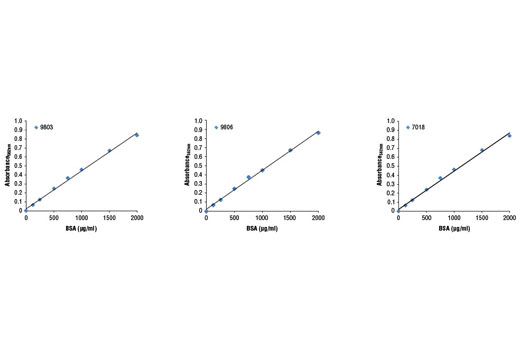
The Coomassie brilliant blue protein assay, commonly known as the Bradford assay 1, is widely used because of its rapid and convenient protocol as well as its relative sensitivity. Pre-diluted Protein Assay Standards:. Most protein assay methods use BSA or immunoglobulin (IgG) as the standard against which the concentration of protein in the sample is determined (Figure 1).
2.1 List of reagents:. To assay protein samples of unknown concentration. Linearization of the Bradford Protein Calibration Graph:.
The Bradford assay is very fast and uses about the same amount of protein as the Lowry assay. Unfortunately, there is a large degree of curvature over a broad range of protein concentrations (Fig. Prepared from the protein and assayed alongside the unknown(s) before the concentration of each unknown is determined based on the standard curve.
The bacterial samples of 30 and 60 minutes from lab 6, and 3. It is a quick and accurate spectroscopic analytical procedure used to measure the concentration of protein in a solution. Without the protein, the solution is red-brown in its acidic solution and when protein binds, the pKa of the dye shifts causing the dye to become blue.
# BE -402B ). It is fairly accurate and samples that are out of range can be retested within minutes. To learn the use of the spectrophotometer.
The Bradford Protein Assay measures protein concentration in a sample. 6) BCA Protein Assay:. In the Bradford assay, we used the dye Coomassie G-250 which binds to proteins mostly at arginine but also at tryptophan, tyrosine, histidine and phenylalanine residues (Olson, 07).
But, i don't understand why it was said that the value closer to 1 is a better indicator to show that my standard curve is good to determine the protein concentration. By measuring absorbance values corresponding to a set of protein standards we can plot a standard curve of absorbance versus amount of protein. Protein assay methods based on similar chemistry have similar protein-to-protein variation.
Bicinchoninic acid assay (Protein. Through this curve you can determine the unknown protein concentration by use of Beer’s Law. Stock solution of Bovine Serum Albumin (BSA):.
Pierce 660nm Protein Assay Reagent, 450mL. Use graph paper or a computer program like Excel to create a scatter plot of the Bradford protein assay data for the BSA parallel dilutions 1-7. A seven-point standard curve for the estimation of target protein amount by the Lowry method.
The standard curve and the unknowns. Linearization of the Bradford Protein Calibration Graph:. The Standard assay is used to measure protein concentrations within a working range of 10 to 00 μg/mL in a 96-well plate.
Bradford and Lowry protein assay reagents results in a. The equation for the curve is y = -4x10-5 x 2 + 0.0115x - 0.0027. Explain why or why not.
The absorbance readings obtained from these solutions are used to construct a graph of absorbance as a function of protein concentration. When you graph your protein assay data in Excel, should you include the absorbance of your unknown BSA sample?. Bovine Serum Albumin (BSA) Set, 7 × 3.5mL, contains.
Diagram of protein assay steps. 0129.7 230 Coomassie (Bradford) Protein Assay Kit. Hydrolysis is probably the most accuratemethod of determining protein concentration followed by amino acid analysis.
A standard protein curve is made to show the relationship of the concentration of the protein to that of the absorbency at 595nm. This graph is called the standard curve for assay, and can be used to convert the absorbance readings for the experimental samples (eggs from duck, chicken 1 (ayam biasa), chicken 2 (ayam kampung), omega 3. Because the color response with coomassie is non-linear with increasing protein concentration, a standard curve must be completed with each assay.
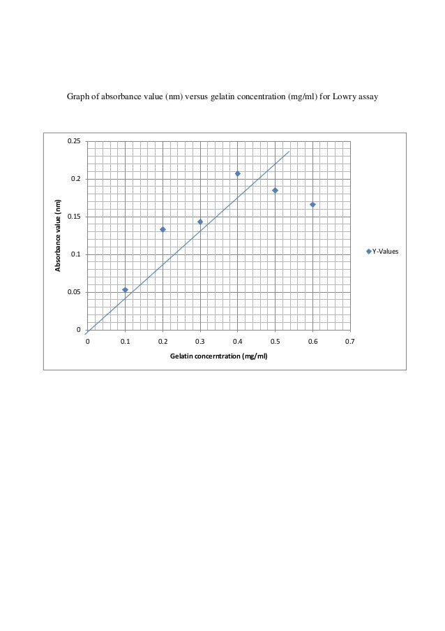
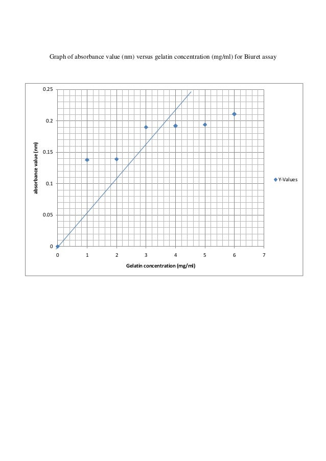
Graph of absorbance value (nm) versus gelatin concentration (mg/ml) for Biuret assay 0.25 0. The Coomassie brilliant blue protein assay, commonly known as the Bradford assay 1, is widely used because of its rapid and convenient protocol as well as its relative sensitivity. -we are going to do the protein assay for three samples:.
The Bradford assay is a colorimetric assay that measures protein concentration. 2 Click twice on the default sheet name in the Prism Navigator tree to rename the sheet. Coomassie Protein Assay Teacher ï book (Cat.
It gives an easy way to estimate the protein concentration of a solution using a standard curve generated by the use of known concentrations of a protein. Create the BSA protein standard curve from your Bradford protein assay data:. The Bradford protein assay was developed by Marion M.
However, if great accuracy is required, prepare the standard curve from a pure sample of the target protein. Biuret Protein Assay Teachers Hand book (Cat. Pierce 660nm Protein Assay Kit, sufficient reagents for 300 test tube or 3000 microplate assays.
A protein assay consists of two main components. This protocol allows the preparation of a nine-point serial dilution of BSA standard in order to obtain a standard curve. + bx + c, where solving for x determines the protein concentration of the sample.
Pierce™ BCA Protein Assay Kit. The absorbance is measured using a spectrophotometer, at the maximum absorbance frequency (A max) of the blue dye (which is 595 nm). Protein concentration is the independent variable (x) and A595 is the dependent variable (y).
If standard (top row) and unknown (bottom row) samples are dispensed and mixed with the same amount of assay reagent, then they are directly comparable. Briefly describe the principles behind the protein assay and their weakness and strengths. Enjoy the videos and music you love, upload original content, and share it all with friends, family, and the world on YouTube.
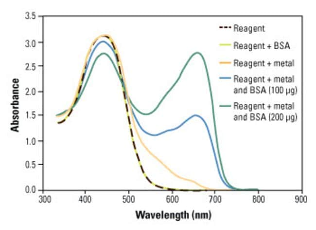
Briefly describe the principles behind the protein assay and their weakness and strengths. If precise quantitation of an unknown protein is required, it is advisable to select a protein. EDTA, Tris, carbohydrates, reducing agents (e.g., DTT, 2-mercaptoethanol), and potassium and.
The absorbance of the BSA protein standards (Tubes 1–5) in each assay. The Bradford protein assay is a dye-binding assay based on the differential color change of a dye in response to various concentrations of protein. Image depicts the screen output from KC3 of a typical standard curve of a Lowry protein assay.
It is a sensitive technique. The Bradford is the reverse:. Like other protein assays, the result of the Bradford assay differs for different proteins.
If the absorbances of the final solutions (green) are identical, then the concentration of the unknown sample is determined to be 1 mg/mL. Add 1 ml of 1 M NaOH with 0.1 ml of diluted sample plus 5 ml of reagent, protein assay 230 Pierce, stirred with a vortex mixer. Absorbance of protein (raw fish) = 0.280 A y = 0.1276 ln (x) + 0.0346 ln x = 0.280 – 0.0346 0.1276 x = 6.84 mg/ml (protein concentration) We have plotted the graph for the standard gelatin and raw fish protein.
µg protein added 0 2.5 5 7.5 10 12.5 15 17.5 25 30 35 2mg/mL BSA (μL) 0 1.25 2.5 3.75 5 6.25 7.5 8.75 10 12.5 15 17.5 H. The data below should not be used as a replacement of a standard curve.
Q Tbn 3aand9gcqpvst Dwjmf0cig7j8wdewswqbb2pje5ywxdeave9j8y8nug44 Usqp Cau

Quantification Of Soluble Or Insoluble Fractions Of Leishmania Parasite Proteins In Microvolume Applications A Simplification To Standard Lowry Assay

Bradford Protein Assay Wikipedia
How To Create A Linear Standard Curve In Excel

G Biosciences Cb X Protein Assay 500 Assay Cb X Protein Assay 500 Assay Fisher Scientific

Sensitive Protein Assay Overcomes All Interfering Agents No Protein To Protein Variation

Protein Assay Lab Vannimah
Q Tbn 3aand9gcqqpwmbrq0qz8qo1g7 7 Umleizjlvestldc3nc1desq2lnbf0a Usqp Cau
Molecular Endocrinology Laboratory Spectrophotometry
Assets Thermofisher Com Tfs Assets Lsg Manuals Man Mod Lowry Protein Asy Ug Pdf
Guide To Enzyme Unit Definitions And Assay Design Biomol Blog Resources Biomol Gmbh Life Science Shop

a Protein Assay Kit Ab Abcam

Nick S Teaching Blog Why Can T I Extrapolate The Bradford Assay Graph If The Beer Lambert Law Applies

Protein Analysis Using Microplate Based Quantification Methods June 28 18
Bradford Protein Assay Calculation Of An Unknown Standard
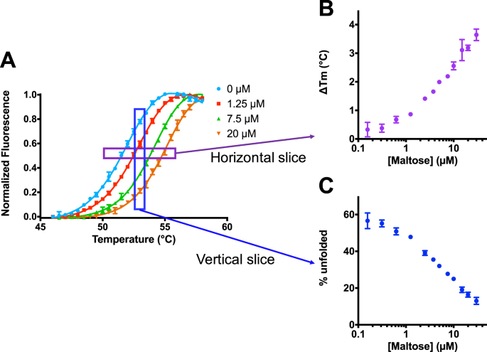
Isothermal Analysis Of Thermofluor Data Can Readily Provide Quantitative Binding Affinities Scientific Reports

Bradford Protein Assay Principle Protocol Calculations Video Lesson Transcript Study Com

Biochemistry Practical 2 Protein Experiment

Protein Assay Lab Report Jane S Ap Bio Webpage
Use The Spectrovis Plus To Study Proteins

Standard Curve Wikipedia
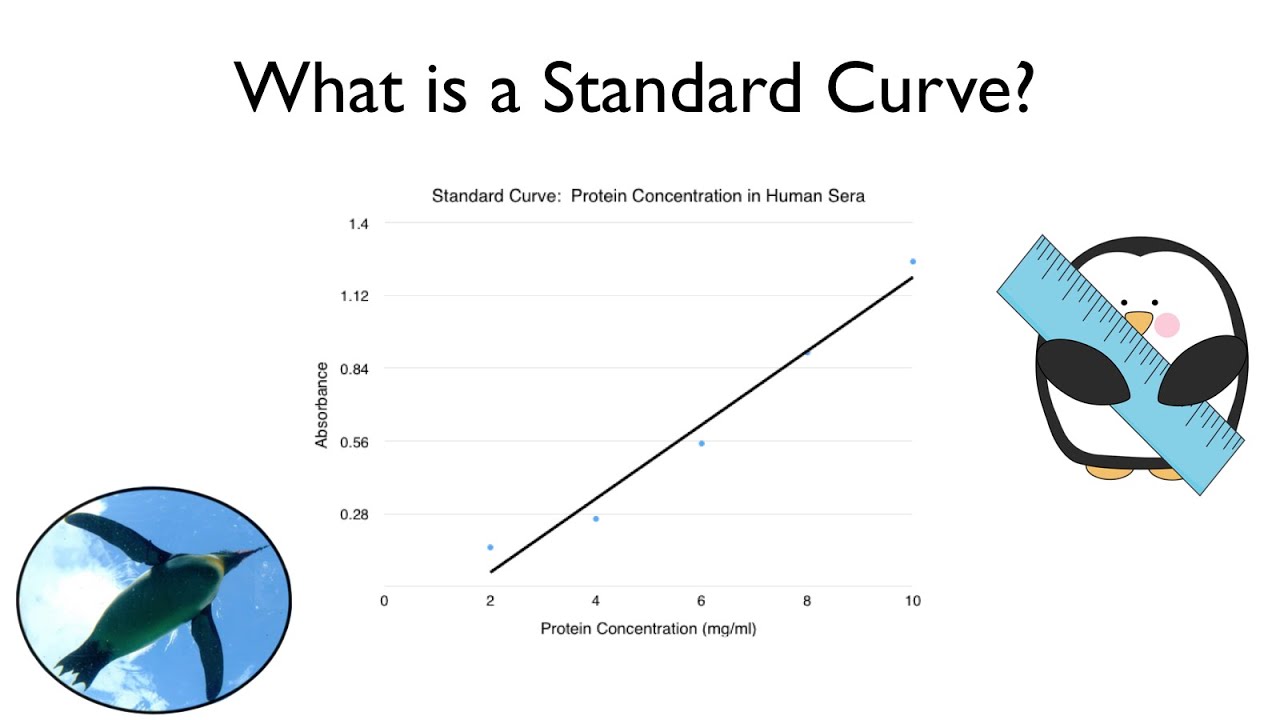
What Is A Standard Curve Youtube
Bradford Protein Assay Calculation Of An Unknown Standard
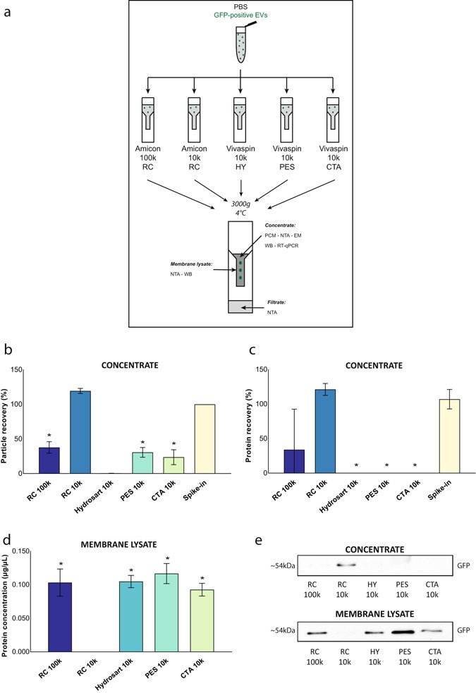
Confounding Factors Of Ultrafiltration And Protein Analysis In Extracellular Vesicle Research Scientific Reports

Assays For Determination Of Protein Concentration Olson 07 Current Protocols In Protein Science Wiley Online Library

B Bsa Calibration Curve This Standard Curve Of Protein Concentration Download Scientific Diagram
Linearization Of The Bradford Protein Assay Protocol

Bradford Protein Assay Lab Adrian Biology

Standard Curve For The Determination Of Protein Concentration By Biuret Download Scientific Diagram

Graph Showing Linear Plot Of Bsa Standards Used In Protein Download Scientific Diagram

Linearization Of The Bradford Protein Assay Protocol
2

Lab 6 Protein Quantification Biology1

Assays For Determination Of Protein Concentration Olson 07 Current Protocols In Protein Science Wiley Online Library
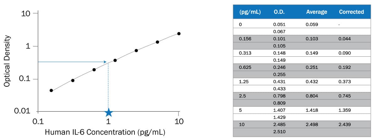
How To Analyze Elisa Data R D Systems

Figure 8 From Linearization Of The Bradford Protein Assay Semantic Scholar

Figure 5 From Linearization Of The Bradford Protein Assay Increases Its Sensitivity Theoretical And Experimental Studies Semantic Scholar

A Analysis Graph Of Total Protein Quantities Of All Groups Detected Download Scientific Diagram
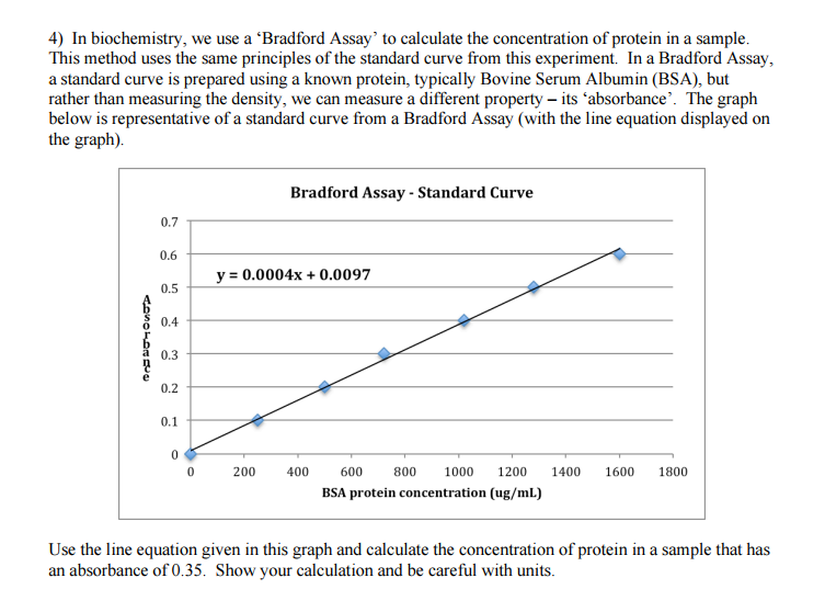
Solved In Biochemistry We Use A Bradford Assay To Calc Chegg Com
Linearization Of The Bradford Protein Assay Protocol
How To Create A Linear Standard Curve In Excel

Protein Assay Writing Assignment Graph Guidelines Youtube

a Protein Assay Kit Ab Abcam

Pierce a Protein Assay Protocol
2

Standard Curve An Overview Sciencedirect Topics

R For Biochemists Let S Analyse A Protein Assay

Standard Curve An Overview Sciencedirect Topics

Standard Curve An Overview Sciencedirect Topics
Setting Up A Colorimetric Assay

Amplite Fluorimetric Fluorescamine Protein Quantitation Kit Blue Fluorescence t Bioquest

a Assay For Protein Determination Nfsc 450
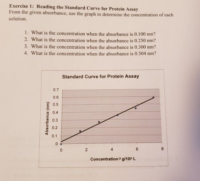
Solved Exercise 1 Reading The Standard Curve For Protein Chegg Com
Solved Bradford Assay Lab Compare Your Protein Concentrat Chegg Com

Protein Analysis Using Microplate Based Quantification Methods June 28 18
a Protein Assay Kit

Bradford Assay Calculations Youtube
Calibration Graph For Protein Using The Bradford Assay For Smartphone Download Scientific Diagram

Prism 3 Calculating Unknown Concentrations Using A Standard Curve Faq 1753 Graphpad

Pdf Linearization Of The Bradford Protein Assay Semantic Scholar

Solved You Are Given 0µl Of A Sample Of Protein A For Chegg Com
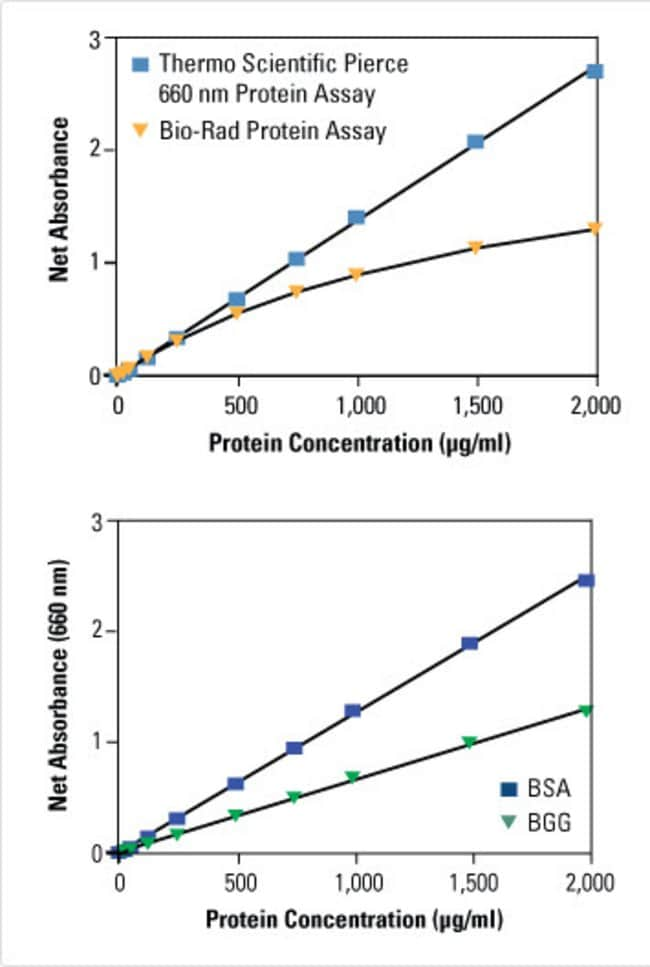
Pierce 660nm Protein Assay Reagent

Bradford Protein Assay Wikipedia
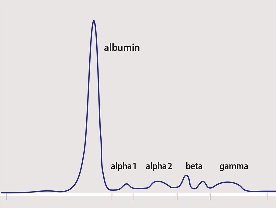
Measuring M Protein Spep Upep Tests Int L Myeloma Fdn

Linear Range Of Protein Quantification For The Lowry And Bradford Download Scientific Diagram

Figure 1 From Linearization Of The Bradford Protein Assay Semantic Scholar

Protein Assay
2

Cb X Protein Assay With Bsa Standard From G Biosciences Biocompare Com
Q Tbn 3aand9gcq8qafndghkgwuy2syn7t3eh3kbhvyjuxtqgeneq4i 2kezwka6 Usqp Cau

User Keyun Wang Notebook Experimental Biological Chemistry I 12 11 07 Openwetware

Protein Measurement Find A Suitable Method Bmg Labtech

Bradford Protein Assay Wikiwand

Team Sdu Denmark Experiments 16 Igem Org
Pierce 660nm Protein Assay Reagent
Cb Protein Assay Protein Quantification Assay Protein Research

In Vitro Protein Releasing Assay Graph Shows The Protein Concentration Download Scientific Diagram
Linearization Of The Bradford Protein Assay Protocol

Responses Of Different Proteins In The Bradford Assay
Http Datasheets Scbt Com Sc 23 Pdf

Calibration Graph For Protein Using The Bradford Assay For Smartphone Download Scientific Diagram

Standard Curve An Overview Sciencedirect Topics
Lowry Vs Biuret Final 1
2
Http Himedialabs Com Td Htbc005 Pdf
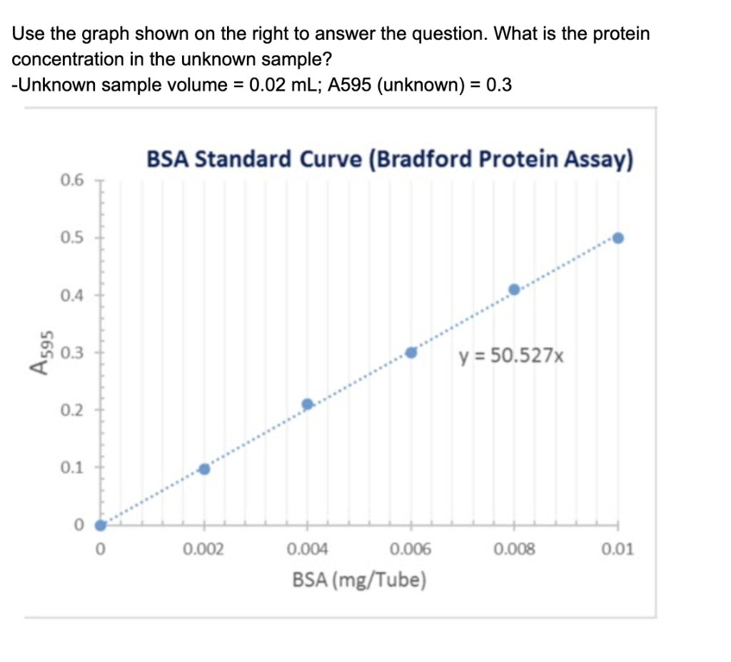
Solved Use The Graph Shown On The Right To Answer The Que Chegg Com

Log Versus Log Scale Plot Of A Standard Curve For Results From 10 Download Scientific Diagram
Q Tbn 3aand9gctwaf5pjnd Ytgtthkdm6yio6vgnvstjzputfjtg4zu9p7nlo15 Usqp Cau
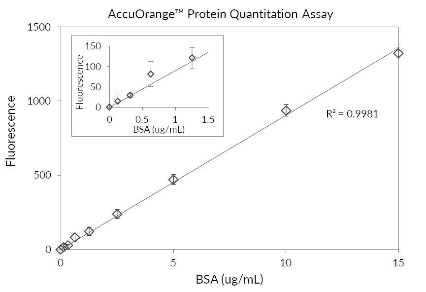
Accuorange Protein Quantitation Kit Biotium
Lowry Vs Biuret Final 1

G Biosciences Protein Assay Cb 500 Assays Protein Assay Cb 500 Assays Fisher Scientific

Linear Range Of Detection And What It Means For Your Quantifications Azure Biosystems

Protein Concentration Using Excel 16 Youtube

R For Biochemists Drawing The Protein Assay With Ggplot

Protein Assay Lab Sudhapetluri

The Use Of Multiplex Platforms For Absolute And Relative Protein Quantification Of Clinical Material Sciencedirect

Bradford Protein Assay Calculation Of An Unknown Standard Protein Bradford Standard
Http Webservices Itcs Umich Edu Drupal Allenlab Sites Webservices Itcs Umich Edu Drupal Allenlab Files Allen lab bca assay protocol Pdf


